


Endothelial microscopy
Endothelial microscopy
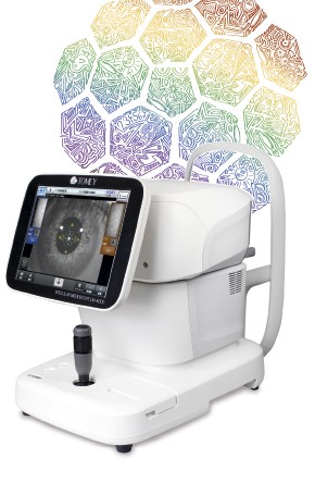
New eye examination – corneal endothelial microscopy using the state-of-the-art TOMEY EM-4000 microscope and digital data processing.
Endothelial microscopy is the quantitative and qualitative evaluation of the eye front transparent layer – corneal – internal cell layer endothelium. For this purpose a cutting edge automated non-contact light-reflecting TOMEY EM-4000 microscope and modern digital data processing are applied.
Advantages of TOMEY EM-4000 microscope:
- Non-contact measurement;
- Auto alignment;
- Measurements are made quickly and accurately (few seconds per one eye);
- 190 x optical magnification;
- Built-in non-contact pachymetry (measurement of the central corneal thickness);
- Measurement of 13 zones of the cornea is possible (1 central+ 6 paracentral+ 6 peripheral).
What does software built-in the microscope identify?
- Density of the endothelial cells (cell number /mm²); the density of endothelial cells of the most people with normal cornea is at least 2000c/mm² and the difference between both eyes is less than 300c/mm²;
- Cell’s polymegathism (variation in cell size) reflected by CV (variation coefficient, norm= 0.22- 0.31); where CV is greater than 0.40, the function of the endothelium can still be adequate, but the endothelium is exposed to a greater risk of damage, in a case of intraocular surgery, or development of glaucoma, diabetes or uveitis or if you wear contact lenses;
- Cell’s pleomorphism (morphology – assessing the percentage of hexagonal cell quantity); an ideal corneal endothelium should consists of hexagonal cells. A normal human cornea should contain ~60% of hexagonal cells;
- Automatically identifies “dark” zones. Dark spots in the endothelium appear both in a case of primary and secondary endotheliopathy – after intraocular surgeries, eye inflammations, glaucoma, wearing contact lenses.
When it is recommended to undergo endothelial microscopy?
- An accurate diagnostics of the endothelium can help to identify not just the reason for changes, but also to plan treatment of frequent eye diseases – glaucoma, uveitis, Fuchs endothelial dystrophy. These diseases can cause changes in the endothelial structure and functionality, which can result in the development of the corneal oedema and vision deterioration. Also wearing contact lenses and intraocular surgeries can cause changes in the endothelium and contribute to the development of oedema in the cornea;
- Where a cataract surgery is planned;
- Where the endothelium exhibits lowered quality indices the surgeon can plan a cataract surgery so that to traumatize the cornea as little as possible during the surgery. Prior to the surgery it is possible to assess the risk of development of a corneal oedema and inform the patient of the risks;
- After surgery to evaluate changes in the condition of the cornea;
- In refractive surgery. Where the endothelial cell density is low, the vision correction with intraocular lens implantation (e.g., Artisan or ICL) is not recommended. Where the surgery has been done, it is advisable to monitor the cell density to make sure that no chronic reduction of cell density is developing, which needs adequate treatment;
- After corneal transplant surgeries. A regular control of the condition of the endothelium after the corneal transplantation is important in monitoring of the transplanted tissues. Sudden reduction of cell density after a corneal transplant (keratoplastic) surgery can evidence of the inflammatory response or immune rejection response / apoptosis, which need immediate treatment. The cell density after transplantation serves as the index of the transplant longevity expectancy;
- In a glaucoma surgery. After glaucoma surgeries, specifically with implantation of filtration elements, the corneal decompensation can also develop. The endothelial cells can be damaged both from the contact of the implant itself with the cornea or as a result of the immune response.
Useful information or when it is allowed /not allowed to perform endothelial microscopy using TOMEY EM-4000
- There are no age restrictions to undergo measurement. Measurement may be done on small children as well;
- The device performs measurement very quickly and painless, during the procedure there are no whatsoever unpleasant sensations, it is not necessary to remain for a long time in a forced position. During examination the patient remains seated at the device with a supported chin;
- The device is not portable, therefore it is impossible to perform measurement for bed-bound patients;
- Measurement can fail, if there is a large corneal oedema or extensive scarring involving corneal opacity, or also if there is an apparent damage of the endothelium, when the cell borders are not traceable at all.