


Degeneration of macula
What is degeneration of macula?
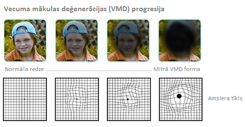
When eye looks directly macula is the central point of retina where the rays of light coming through cornea and lens are bent. Similarly as with the lens in photo camera image gets on retina through the lens that resembles opening of the camera. If changes have occurred in macula, central part of the image is lost, as if a blurred, blended patch would be in front of it. The rest of the image could remain clear.
What causes degeneration of macula?
Degeneration of macula is caused by changes or damages in macula. The eye is still able to see side objects very clearly but the peripheral vision is usually disturbed. Due to this reason degeneration of macula alone is not able to cause blindness. Still reading or precise work from the close distance is troubled or impossible.
Retina is a delicate and sensitive layer of tissues covering internal wall of the eyeball. Macula is just a small space located in the centre of retina that can be compared to the big letter "O" in the newspaper. Our "straight forward" vision that we use for reading and other delicate works is impossible without this small area. Although degeneration of macula is most frequently absorbed for elderly patients not always the loss of central vision takes place only because eye is aging. Sometimes eye traumas, infections or inflammations cause degeneration of macula.
What are the forms of macular degeneration?
Most common form of the macular degeneration is involutionary degeneration of macula. This form is established in approximately 70% cases and related to the aging process of the eye. It is caused by the disintegration or separation of the tissues of macula, as they become thinner, more delicate and fragile.
Exudative degeneration of macula can be established in approximately 10% of the cases. Usually a thin layer of tissues protects macula, separating it from the back of the eye with richly covered, very delicate blood vessels. Sometimes those blood vessels burst or exude sap (bleeds), as a result scar tissues are formed. Frequently, a new blood vessels that eye does not need form and grow into those scar tissues. Those new blood vessels are especially delicate and sensitive they easily burst and exude sap. Blood and exuding of sap destroys macula and causes further formation of scars. Vision is distorted and blurred and dense scar tissues block central vision of the eye.
There are also forms of macular degeneration that are found in young patients (teenager/adolescent macular degeneration) that are not caused by the aging process. Sometimes trauma, infection or inflammation causes damage of the delicate tissues of macula.
What are the symptoms of macular degeneration?
If macular degeneration has happened only in one eye, then in the beginning it is hart to notice it, if the second eye is in a good condition. Frequently it is usually this case and changes in vision are diagnosed after a large amount of time. Symptoms of macular degeneration can be different to every patient. Sometimes only one eye loses vision while in the other eye good vision remains for years. If both eyes are affected, then reading and performing of delicate works becomes especially inconvenient.
Degeneration of macula does not cause blindness on its own. As the side vision is not usually affected, most of the patients can take care of themselves sufficiently well.
One of the symptoms of macular degeneration could be dull image of colour, letters look blurred while reading, straight lines look bended and sometimes central part of the image looks more distorted than the rest of it. Dark or blurred field is seen in the centre of image.
How to diagnose macular degeneration?
Many patients are not aware that they have been affected by macular degeneration until severe vision changes appear. An eye doctor can recognize macular degeneration at an early stage.
During the visit, the doctor clarifies complaints of the patient, reviews data on the patient’s medical history, as well as the family health history. The doctor conducts a complete eye exam and vision health diagnosis, during which the doctor determines the visual functions, as well as testing of all the eye structures.
The doctor conducts an examination - OCT (optical coherence tomography) that specifies the form and extent of the disease. It is a painless, noninvasive imaging test, displaying images of the retina in a high-resolution version.
In addition, fluorescent angiography can be done. During this test, a colored dye called fluorescin is injected into a vein in your arm that travels to and highlights the blood vessels in your retina and a special camera takes a picture of the retina and the blood vessels.
Early diagnosis can prevent further changes in the macula or help choose ophthalmic optics to improve vision.
How macular degeneration is cured?
Early stages of the exudative degeneration of macula can be treated with the ophthalmic laser. Beam of the laser cauterize exuding membranes and destroys newly formed blood vessels, thus reducing further changes of the loss of vision that can be caused by the progressive scaring of macula and surrounding retina.
Age-related macular degeneration (AMD)
The age-related macular degeneration (AMD) is the leading type of the macular degeneration associated with aging of the eye, causing permanent damage to the back of the eye.
Usually, AMD affects both eyes but these signs can initially appear in one eye. If the age-related macular degeneration progresses in the late stages of the disease, a severe and permanent loss of vision may develop.
Most commonly, loss of vision occurs gradually and damages the central portion of vision, and this means that each eye of the patient is still able to see from the side.
http://cv-eye.com/procedures/retina/macular-degeneration/
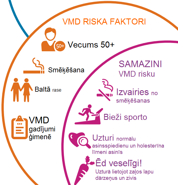
Risk factors
AMD is a common cause of poor vision for older people – the greater the age, the greater the risk of the disease. The age-related macular degeneration is an inherited eye disease. Relatives of AMD patients are at an increased risk. Also, smoking and diet that lack certain important nutrients greatly increase the risk of degeneration.
Types of AMD
Early AMD - the metabolic end products accumulate under the macula, create deposits that are called drusen. You may not complain of vision impairment but at this stage of the disease, the change of diet and lifestyle may help slow down the development of the disease.
Advanced AMD - the disease progresses, there is damage to the eyesight. There are two advanced types of AMD:
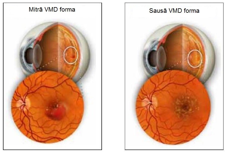
1. Dry AMD - most commonly develops slowly and can advance over the course of several years. In the macula, cells die in small areas, images appear blurry and pale, image details are lost.
2. Wet AMD - new, pathological blood vessels grow from the choroidal layer (back of the eye) inside the macula or under it. These blood vessels are bleeding and secreting. On the macula or around it, scar tissues are formed. Wet-type AMD develops more rapidly and an evident visual impairment is felt within weeks, days or even hours. The signs are as follows: deformation of visual objects, blank or blurred spots in the portion of the central vision. Two-thirds of cases of the advanced AMD account for the wet age-related macular degeneration.
http://healthlifemedia.com/healthy/wp-content/uploads/2016/12/Wet-and-Dry-AMD.jpg
Early detection of the disease
If you are 50 years of age, it is valuable to plan visits to the eye doctor every year or every other year in order to detect early AMD when vision is not yet damaged. Early detection of the age-related macular degeneration is more likely to delay vision impairment.
You can monitor the age-related macular degeneration through carrying out the Amsler grid test on a daily basis, using adequate spectacles for reading if you need them. If you detect vision changes, immediately contact the eye doctor.
If you have AMD and no vision complaints, using the Amsler's grid test on a daily basis can replace regular visits to your eye doctor.
If you have been diagnosed with AMD, it is important together with the eye doctor to understand the disease, its development, and the treatment plan.
Treatment of wet AMD
Treatment of wet AMD aims to prevent deterioration in the eyesight. Early treatment of the disease may better maintain good vision. Treatment includes:
• Anti-VEGF (vascular endothelial growth factor) drugs that reduce a new secretion of blood vessels or bleeding in the macula.
• Photodynamic treatment – light-sensitive drugs and laser treatment eliminate the pathological blood vessels.
• Laser photocoagulation - a laser beam usage near the pathological blood vessels.
Early and dry treatment of AMD
Treatment involves a diet change, quitting smoking and, in some cases, the use of vitamin supplements.
What can I do?
• If you smoke, quit it now. Smoking greatly increases the risk of development of AMD and the advanced stage of the AMD disease, the advanced AMD can begin sooner.
• If you are 50 years of age, visit the eye doctor every year or every two years when it is possible to recognize AMD in the early stage.
• Tell the eye doctor if your relative has AMD.
• Do the Amsler grid test every day to follow the early signs of AMD. If you have a sudden change in vision, immediately make an appointment with the eye doctor.
• The daily use of fruits, green, leafy vegetables twice or thrice a week, fish, a handful of nuts per week, low glycemic (low-GI) index carbohydrates reducing fats and oils will contribute to your health.
• Discuss with your doctor whether using dietary supplements of vitamins will be beneficial to your health.
• Maintain average weight through doing regular exercises.
Given that AMD is a hereditary disease, it is possible to carry out a genetic test (including young people) to identify potential lifetime risks of having AMD.